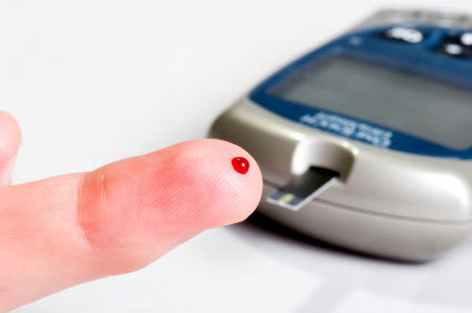
Alterações cerebrais são evidentes no Diabetes Mellitus

O Diabetes Mellitus (DM) acomete uma grande parte da população mundial. A fisiopatologia dessa doença é principalmente lincada à destruição das células Beta pancreáticas. Entretanto, estudos recentes mostram que o DM também acomete o sistema nervoso central. Pacientes que sofrem de DM apresentam prejuízo cognitivo e uma maior probabilidade de terem transtornos psiquiátricos, incluindo a depressão. Muitos desses estudos foram realizados pela unidade de pesquisa em depressão do laboratório de Neurociências da UNESC. Um estudo aceito recentemente para publicação no periódico Current Neurovascular Research em parceria com o Laboratório de Fisiopatologia Experimental da UNESC revelou que o DM induzido experimentalmente levou a alterações importantes em proteínas cerebrais envolvidas com a plasticidade celular. O estudo fez parta da tese de doutorado de Maria Augusta B. dos Santos.
O resumo da publicação pode ser visualizado a seguir:
Curr Neurovasc Res. 2016 Feb 18.
Antioxidant therapy alters brain MAPK-JNK and BDNF signaling pathways in experimental Diabetes Mellitus.
Réus GZ, Dos Santos MA, Abelaira HM, Maciel AL, Arent CO, Matias BI, Bruchchen L, Ignácio ZM, Michels M, Dal-Pizzol F, Carvalho AF, Zugno AI, Quevedo J.
Abstract
This study was designed to investigate the effects of treatment with the antioxidants N-acetylcysteine (NAC) and deferoxamine (DFX) in intracellular pathways in the brain of diabetic rats. To conduct this study we induced diabetes in Wistar rats with a single injection of alloxan, and afterwards rats were treated with NAC or DFX for 14 days. Following treatment completion, the immunocontent of c-Jun N-terminal kinase (JNK), mitogen-activated protein kinase-38 (MAPK38), brain-derived neurotrophic factor (BDNF), and protein kinases A and C (PKA and PKC) were determined in the prefrontal cortex (PFC), hippocampus, amygdala and nucleus accumbens (NAc). DFX treatment increased JNK content in the PFC and NAc of diabetic rats. In the amygdala, JNK was increased in diabetics treated with saline or NAC. MAPK38 was decreased in the PFC of control and in diabetic rats treated with NAC or DFX; and in the NAc in all groups. PKA was decreased in the PFC with DFX treatment. In the amygdala, PKA content was increased in diabetic rats treated with either saline or NAC, compared to controls; and it was decreased in either NAC or DFX-treated groups, compared to saline-treated diabetic animals. In the NAc, PKA was increased in NAC-treated diabetic rats. PKC was increased in the amygdala of NAC-treated diabetic rats. In the PFC, the BDNF levels were decreased following treatment with DFX in diabetic rats. In the hippocampus of diabetic rats the BDNF levels were decreased. However, treatment with DFX reversed this effect. In the amygdala the BDNF increased with DFX in non-diabetic rats. In the NAc DFX treatment increased the BDNF levels in diabetic rats. In conclusion, both diabetes and treatment with antioxidants were able to alter intracellular pathways involved in the regulation of cell survival in a brain area and treatment-dependent fashion.
Mais informações: http://www.ncbi.nlm.nih.gov/pubmed/26891662
01 de março de 2016 às 21:03
 Newsletter
  RSS
RSS